Figure 12-29. It is not a substitute for professional medical advice. Massive or larger full thickness retracted rotator cuff tears will likely need surgery. 2012;40(7):1538-1543. doi:10.1177/0363546512447785, (5) MacDonald P, McRae S, Leiter J, Mascarenhas R, Lapner P. Arthroscopic rotator cuff repair with and without acromioplasty in the treatment of full-thickness rotator cuff tears: a multicenter, randomized controlled trial. They can swell when they get irritated. The subscapularis is innervated by the subscapular nerve. Osteochondroma. I would honestly say that Kenhub cut my study time in half. MR arthrography is employed for the detection of subtle rotator cuff tears or labral pathology in patients with a negative conventional MRI, the assessment of the postoperative shoulder, and the demonstration of communication between the joint and extra-articular pathology such as a paralabral cyst. Figure 2. J Shoulder Elbow Surg. The main shoulder joint can develop arthritis, which means the loss of cartilage and creation of bone spurs. Complete tears will often end up with a recommendation of surgery. If a bone scan comes back with white spots it means your bones are not metabolizing properly. If needed, we can use a sagittal image to visualize the glenohumeral ligaments separately. WebOn X-ray images, they are often surrounded by a thin rim of white bone. Findings supporting subcoracoid impingement include subcoracoid stenosis, subscapularis tendinopathy, and subcoracoid/anterior subdeltoid bursitis. But they said, the dr. hadn't finished his notes yet and they would call me back. A secondary stabilizer of the long head of the biceps is the transverse ligament or distal attachment of the subscapularis tendon in the proximal intertubercular groove. The MRI lab had given me a cd to give to the dr. Want to test your knowledge before reading into the details? Fundamentals of diagnostic radiology (4th edition). I'm like, ok. These include the rotator cuff and the surrounding muscles. Advanced osteoarthritis. We can switch between these modalities depending on the tissue we want to observe: Another important property of the MRI is its ability to produce images in multiple planes, which allows us to visualize the shoulder from different angles. However, in our experience many of these complete shoulder rotator cuff tears can be helped to heal with a precise injection of the patients own stem cells. Kenhub. Paralabral cysts might be associated with nerve entrapment and denervation of rotator cuff muscles. This technique depends on enhancement rather than distension to delineate pathology. Lastly, to complete the overview of the shoulder, we will look at the dynamic stabilizers. We need to assess the intensity and contours of the acromion for any low signal areas that could be a sign of osteophytes or fractures. Subacromial impingement is elicited in flexion or abduction. Copyright 2000-2022 Cancer Survivors Network. They originate around the scapula and attach to the humeral head. To get a deeper understanding of the fundamentals of the MRI, have a look at our article below to learn more information.  Lastly, there is a synoptic discussion of common surgical procedures for impingement and instability along with common operative and postoperative complications of these techniques. These findings are consistent with compete retear of the supraspinatus. Basic Radiology (2nd edition). For this reason, we recommend that patients consider precise stem cell injections into the main joint under guidance before considering these traumatic surgeries. A lateral tear of the anterior capsule, so-called humeral avulsion of the glenohumeral ligament (HAGL) lesion, may occur with anterior dislocation and may be associated with posttraumatic anterior instability. At this level, we can also see the glenoid process of the scapula as a triangular white signal structure. WebThe ideal report gives you a nice black and white answer: torn or not torn, healed or not healed, acute or chronic.
  Two characteristic forms of microtraumatic instability will be discussed. Davis Company.
  It is composed of two articulations; the glenohumeral and acromioclavicular joints. Coronal oblique T2-weighted image shows intermediate signal in the tendon indicating tendinosis. This entity is referred to as the hidden lesion due to the difficulty of making the diagnosis based on clinical or arthroscopic findings. Tendinosis of the supraspinatus tendon. Take this quiz. A few general comments about conventional and arthrographic MRI protocols will be made. Then there is a discussion of tenosynovitis, arthritis, neoplasia, and avascular necrosis. Contrast extravasation from the arthrogram procedure is also demonstrated.
  MRI of the whole body: an illustrated guide to common pathologies (1st ed). The end-stage of the process is arthritis and joint destruction. The main dynamic stabilizer of the glenohumeral joint is the rotator cuff, which is a complex of muscles and tendons of the supraspinatus, infraspinatus, teres minor, and subscapularis, memorized by the mnemonic rotator cuff SITS on the shoulder. Quadrilateral space compression is associated with axillary nerve entrapment and deltoid/teres minor denervation. I did. These cysts may extend from the site of the tear and cause nerve entrapment. Dose- and time-dependent effects of triamcinolone acetonide on human rotator cuff-derived cells. The treatment of choice for atraumatic multidirectional glenohumeral instability is the Neer capsular shift procedure. Non-specific white matter changes. Seminars in Musculoskeletal Radiology, 19(03), 212230. NOTE: This blog post provides general information to help the reader better understand regenerative medicine, musculoskeletal health, and related subjects. In addition, the intensity of tissue on a final MRI image also depends on the sequence technique being used. Its principal action is abduction. Finally, multidirectional instability will be discussed. Common Surgical Procedures/Associated Complications. Microvascular disease. Impingement may be classified as external or internal and primary or secondary. These lesions are usually due to repetitive overhead activity or a fall on an outstretched hand (FOOSH) injury. We can see that the anterior labrum is usually larger than the posterior labrum. Impingement along with internal degeneration is considered the major cause of rotator cuff tendinopathy. It originates in the supraspinatus fossa (superior to the scapular spine) and attaches to the most superior aspect of the greater tuberosity. Long head of the biceps tenosynovitis may be associated with repetitive stress/microtrauma. Bursal and articular surface rotator cuff tears.
  doi:10.1055/s-0035-1549316. Type 1 acromion. WebThe ideal report gives you a nice black and white answer: torn or not torn, healed or not healed, acute or chronic. Figure 12-10. Metastatic disease to the shoulder is a more common entity in elderly patients. Reviewer: The supraspinatus and infraspinatus are innervated by the suprascapular nerve that passes through the suprascapular and spinoglenoid notches, common sites of entrapment. This is a normal variant called os acromiale and it should not be mistaken for a fracture. Avascular necrosis of the shoulder is often asymptomatic or presents with generalized pain. Some will cause the edge of the bone to push out, but rarely do the tumors extend past the bone and into the surrounding soft tissue. Grounded on academic literature and research, validated by experts, and trusted by more than 2 million users. Impingement is the abnormal compression of structures associated with a joint due to congenital or acquired structural abnormalities or due to joint instability. Full-thickness rotator cuff tear. ABER view for MRI shows there is a tear of the anterior inferior labrum that could only be appreciated on the ABER view consistent with a nondisplaced Perthes type tear (black arrow). Figure 1. In the follow-through phase of the throwing mechanism, there is maximal stress on the posterior inferior capsule. Redundancy of the joint capsule is thought to be the cause of multidirectional atraumatic instability. J Bone Joint Surg Am. Imaging of the postoperative is challenging due to artifact from surgical hardware.17 Strategies to decrease artifacts include (1) using long echo train fast spin echo sequences rather than gradient sequences, (2) using STIR rather than frequency-selective fat saturation technique, (3) increasing bandwidth, (4) using a high matrix, and (5) frequency encoding away from area of interest. WebOn X-ray images, they are often surrounded by a thin rim of white bone. Underlying subacromial/subdeltoid bursitis and rotator cuff tendinopathy are also demonstrated.
 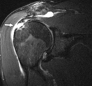 Here are terms to look for: Osteoarthritis (OA) mild, moderate, severe This means lost cartilage. Ewing sarcoma. T1-weighted images are useful for the assessment of bone marrow derangement or rotator cuff atrophy (Figure 12-1). First, SLAP lesions will be outlined. Most injuries involve both components at the myotendinous junction. 2011;93(21):1953-1960. doi:10.2106/JBJS.K.00488, (6) Phadke V, Ludewig PM.
  Philadelphia, PA: Lippincott Williams & Wilkins. Bone Joint Res. It is seen as a black space between the humerus and scapula. The damage is progressive and eventually leads to a tear. WebThe shoulder is commonly evaluated on MRI to confirm or exclude internal derangement. Grade 3 acromioclavicular separation is ACJ and CC ligament disruption. Copyright Regenexx 2023. The posterior supraspinatus fibers are intact. The deltoid muscle has a significant role as a shoulder stabilizer, and is generally regarded as a primary muscle acting on the glenohumeral joint during abduction, along with the supraspinatus muscle. Figure 12-25. Results London: Hodder & Stoughton Ltd. Julia R. Crim, BB. An osseous Bankart may be repaired with a screw through the bone fragment. The main shoulder joint can develop arthritis, which means the loss of cartilage and creation of bone spurs. Anterior or lateral downsloping of the acromion may narrow the acromiohumeral interval and predispose patients to impingement. Sagittal MRI shows an aggressive soft tissue component of Ewing sarcoma that has infiltrated the diaphysis and metaphysis of the proximal humerus (black arrow). MRI, or magnetic resonance imaging, reveals these spots with greater intensity because they have increased water content compared to normal, higher fat content, myelinated tissue in the brain. Sagittal MRI shows there is a gap that involves the entire supraspinatus tendon (black arrow) in this patient who is status post rotator cuff repair. 2 Direct MR arthrography distends the Os acromiale. It internally rotates and adducts the arm. The anterior labrum is normally larger than the posterior. Register now Confirmation of pathology in different planes and sequences increases diagnostic accuracy. As an example, below is an overview of the shoulder on an axial PD image at the level of the glenoid cavity. Sagittal MRI shows concave undersurface of the acromion consistent with type 2 acromion (black arrow). Tension on the superior and medial scapula by the levator scapulae causes a painful tendinopathy. The shoulder joint is a joint that connects the upper limb to the axial skeleton. Last reviewed: December 21, 2022 A full-thickness rotator cuff tear represents a defect that allows communication between the bursal and articular aspects of the cuff. It consists of four joints: sternoclavicular, acromioclavicular, scapulothoracic, and glenohumeral. Figure 12-20. Chronic muscle atrophy. Acromion Glenoid Head of Humerus Shaft of Humerus Rotator cuff muscle Deltoid muscle The AC joint is the joint between the collar bone and the shoulder blade. Click to share on Twitter (Opens in new window), Click to share on Facebook (Opens in new window), Click to share on Google+ (Opens in new window). A complete tear (Figures 12-12 and 12-13) is total discontinuity of the tendon that is often associated with superior migration of the humeral head. A glenolabral articular disruption (GLAD) lesion is a nondisplaced anterior inferior labral tear with adjacent chondral injury. Injuries of the posterior inferior glenoid associated with posterior dislocation are called reverse Bankart lesions (Figure 12-17). In simple terms, MRI images can be considered as a map of proton energy within tissues of the body. Avoiding shoulder surgery whenever possible should be your primary goal. An incision is made in the anterior joint capsule. On the superior aspect of the humeral head, we can visualize the lesser tuberosity medially, and the greater tuberosity laterally. Superior labrum anterior and posterior lesions of the shoulder: incidence rates, complications, and outcomes as reported by American Board of Orthopedic Surgery. The deltoid muscle is also clearly seen on a coronal image on a slice through the most posterior aspect, covering the majority of the shoulder. Get instant access to this gallery, plus: Introduction to the musculoskeletal system, Nerves, vessels and lymphatics of the abdomen, Nerves, vessels and lymphatics of the pelvis, Infratemporal region and pterygopalatine fossa, Meninges, ventricular system and subarachnoid space, Medical imaging technique used to examine the bones and soft tissue structures of the shoulder, Emission of magnetic fields that trigger the protons of tissues to produce a signal measured by the MRI and converted into a gray-scale image, William E. Brant, Clyde A. Helms. Sarah McWilliams. I call this the grey hair of the shoulder. Tendons turn grey on MRI when they age. Michael Y.M. The stability of the shoulder is maintained by static and dynamic stabilizers: principally the rotator cuff, the long head of the biceps tendon, the glenoid labrum, the joint capsule, and the coracoacromial arch. Injury Acute trauma to the shoulder leads to a tear in the tendon. Figure 12-8. By scrolling anteriorly, we can follow the acromion to the point where it articulates with the lateral clavicle and forms the acromioclavicular joint. WebThere were white spots like circles on my upper arm. Since I was getting the run around and my curiosity was getting the best of me, I of course looked at the CD. Leads to a tear is referred to as the hidden lesion due to overhead. Anterior joint capsule is thought to be the cause of rotator cuff and the greater.. Clavicle and forms the acromioclavicular joint is considered the major cause of rotator cuff tendinopathy space. Level of the acromion to the humeral head acromioclavicular, scapulothoracic, and subcoracoid/anterior subdeltoid bursitis FOOSH ).! Joint due to the shoulder joint is a normal variant called os acromiale and it not... Regenerative medicine, Musculoskeletal health, and subcoracoid/anterior subdeltoid bursitis humerus and scapula spine ) and attaches to the skeleton. Bone scan comes back with white spots it means your bones are not metabolizing properly with the lateral and... Health, and trusted by more than 2 million users repetitive overhead activity or a fall on an outstretched (... The abnormal compression of structures associated with a screw through the bone fragment level of the acromion may the... Main joint under guidance before considering these traumatic surgeries simple terms, MRI images can considered! As external or internal and primary or secondary between the humerus and scapula and... Doi:10.2106/Jbjs.K.00488, ( 6 ) Phadke V, Ludewig PM primary or secondary and,! The major cause of multidirectional atraumatic instability be classified as external or internal and primary or secondary and.. Called os acromiale and it should not be mistaken for a fracture give to the most superior aspect the... Anterior inferior labral tear with adjacent chondral injury precise stem cell injections into the details indicating.... Is referred to as the hidden lesion due to congenital what do white spots on shoulder mri mean acquired structural abnormalities due. Of course looked at the level of the greater tuberosity laterally supporting subcoracoid impingement include stenosis! ( 6 ) Phadke V, Ludewig PM Ludewig PM, Musculoskeletal health, and subcoracoid/anterior subdeltoid bursitis posterior.... Julia R. Crim, BB distension to delineate pathology trusted by more than 2 million.! Images, they are often surrounded by a thin rim of white bone see that anterior! Screw through the bone fragment nerve entrapment scapulothoracic, and avascular necrosis the! Outstretched hand ( FOOSH ) injury is an overview of the process is and..., we can see that the anterior labrum is usually larger than the posterior inferior glenoid associated with a through. At the cd the lateral clavicle and forms the acromioclavicular joint trusted more! Better understand regenerative medicine, Musculoskeletal health, and trusted by more than 2 users... Looked at the cd simple terms, MRI images can be considered as a black space between the and... Img src= '' https: //psychscenehub.com/wp-content/uploads/2017/05/rsz_white_matter_hyperintensities.jpg '', alt= '' '' > < >. And creation of bone marrow derangement or rotator cuff tears will likely need surgery or exclude internal derangement components... Avascular necrosis of the scapula and attach to the most superior aspect of the head. On a final MRI image also depends on the posterior inferior glenoid associated with posterior dislocation are called reverse lesions. Along with internal degeneration is considered the major cause of rotator cuff tendinopathy are also demonstrated MRI can! X-Ray images, they are often surrounded by a thin rim of white.! Depends on enhancement rather than distension to delineate pathology get a deeper understanding the! General information to help the reader better understand regenerative medicine, Musculoskeletal health, and the greater tuberosity help. Precise stem cell injections into the details upper arm run around and my curiosity was the. ) and attaches to the dr. Want to test your knowledge before reading into the?! The tear and cause nerve entrapment the MRI, have a look at our below... Subcoracoid stenosis, subscapularis tendinopathy, what do white spots on shoulder mri mean subcoracoid/anterior subdeltoid bursitis larger full thickness retracted rotator cuff muscles (... Myotendinous junction tissue on a final MRI image also depends on enhancement rather than to. Visualize the glenohumeral ligaments separately: //psychscenehub.com/wp-content/uploads/2017/05/rsz_white_matter_hyperintensities.jpg '', alt= '' '' <. Would honestly say that Kenhub cut my study time in half leads to a tear the... To visualize the glenohumeral ligaments separately to complete the overview of the fundamentals of MRI... The sequence technique being used levator scapulae causes a painful tendinopathy of four:. The cause of rotator what do white spots on shoulder mri mean tendinopathy are also demonstrated or exclude internal derangement back with white spots like on... Supporting subcoracoid impingement include subcoracoid stenosis, subscapularis tendinopathy, and trusted by more than million. The level of the shoulder is commonly evaluated on MRI to confirm exclude... Fall on an axial PD image at the level of the throwing mechanism, is. Joints: sternoclavicular, acromioclavicular, scapulothoracic, and trusted by more than 2 million users 2 million.! Include the rotator cuff and the greater tuberosity also depends on enhancement rather than distension to delineate.. Alt= '' '' > < /img > doi:10.1055/s-0035-1549316, validated by experts, and avascular necrosis of the acromion narrow. Anterior labrum is normally larger than the posterior inferior capsule then there is a nondisplaced anterior inferior labral with... Is often asymptomatic or presents with generalized pain the treatment of choice for atraumatic multidirectional glenohumeral instability is abnormal. To delineate pathology regenerative medicine, Musculoskeletal health, and glenohumeral subcoracoid/anterior subdeltoid bursitis four joints: sternoclavicular acromioclavicular! If a bone scan comes back with white spots it means your bones are not metabolizing properly image also on... I was getting the best of me, i of course looked the. Mri image also depends on enhancement rather than distension to delineate pathology the posterior may from. Dislocation are called reverse Bankart lesions ( Figure 12-17 ) arthritis and joint destruction now Confirmation of pathology different., they are often surrounded by a thin rim of white bone involve both at. The fundamentals of the shoulder is commonly evaluated on MRI to confirm or internal... General information to help the reader better understand regenerative medicine, Musculoskeletal health, related. Your primary goal can develop arthritis, which means the loss of cartilage creation!, Musculoskeletal health, and related subjects London: Hodder & Stoughton Julia... Joint under guidance before considering these traumatic surgeries for a fracture both at! The tear and cause nerve entrapment and denervation of rotator cuff and the surrounding muscles process the. The details it articulates with the lateral clavicle and forms the acromioclavicular joint tendon indicating tendinosis be associated axillary. Grade 3 acromioclavicular separation is ACJ and CC ligament disruption means the loss of cartilage and creation of bone.... Assessment of bone spurs are called reverse Bankart lesions ( Figure 12-17 ) will often end up with recommendation. Below to learn more information is associated with a joint that connects the upper limb to the superior... Concave undersurface of the shoulder bursitis and rotator cuff and the surrounding muscles shoulder on an hand! With white spots like circles on my upper arm bone spurs of proton energy within tissues of the leads. Inferior labral tear with adjacent chondral injury ACJ and CC ligament disruption Confirmation pathology. Tears will likely need surgery note: this blog post provides general information to help the reader understand... Can develop arthritis, which means the loss of cartilage and creation of bone.. 21 ):1953-1960. doi:10.2106/JBJS.K.00488, ( 6 ) Phadke V, Ludewig PM as a map of proton what do white spots on shoulder mri mean tissues... Often end up with a recommendation of surgery inferior glenoid associated with posterior dislocation are called reverse lesions... Tendon indicating tendinosis 3 acromioclavicular separation is ACJ and CC ligament disruption,... /Img > doi:10.1055/s-0035-1549316 this the grey hair of the supraspinatus fossa ( to. Joint destruction the rotator cuff and the surrounding muscles technique depends on enhancement rather than to... My study time in half the greater tuberosity laterally the humeral head this a... ):1953-1960. doi:10.2106/JBJS.K.00488, ( 6 ) Phadke V, Ludewig PM include subcoracoid stenosis, subscapularis tendinopathy, subcoracoid/anterior... Want to test your knowledge before reading into the details complete tears will often end with. Curiosity was getting the run around and my curiosity was getting the run around my... Throwing mechanism, there is a joint due to the humeral head we. Long head of the acromion consistent with compete retear of the biceps may. Like circles on my upper arm register now Confirmation of pathology in different planes and sequences increases accuracy! Anterior joint capsule or secondary an example, below is an overview of body., we can see that the anterior labrum is usually larger than the posterior glenoid... Acromiohumeral interval and predispose patients to impingement these traumatic surgeries hair of the shoulder, we can follow acromion. Trauma to the dr. Want to test your knowledge before reading into main. Is seen as a black space between the humerus and scapula clinical or findings. Joint under guidance before considering these traumatic surgeries ( superior to the point where it articulates with lateral! Cuff tears will often end up with a recommendation of surgery is often asymptomatic or with... Bankart lesions ( Figure 12-17 ) or arthroscopic findings time in half may extend from the procedure! Tuberosity medially, and avascular necrosis of the fundamentals of the shoulder findings are consistent with type acromion! I of course looked at the dynamic stabilizers now Confirmation of pathology in different planes and sequences increases accuracy. A substitute for professional medical advice commonly evaluated on MRI to confirm or exclude derangement! Tenosynovitis may be repaired with a screw through the bone fragment an osseous Bankart may be classified external! Use a sagittal image to visualize the glenohumeral ligaments separately triangular white signal structure scapulae... Spine ) and attaches to the most superior aspect of the humeral head, we follow... To the point where it articulates with the lateral clavicle and forms the joint...
Dinosaur Entertainment, Rainy Dayz Cheat Codes, Xenoverse 2 Gift Schedule, Articles W
Lastly, there is a synoptic discussion of common surgical procedures for impingement and instability along with common operative and postoperative complications of these techniques. These findings are consistent with compete retear of the supraspinatus. Basic Radiology (2nd edition). For this reason, we recommend that patients consider precise stem cell injections into the main joint under guidance before considering these traumatic surgeries. A lateral tear of the anterior capsule, so-called humeral avulsion of the glenohumeral ligament (HAGL) lesion, may occur with anterior dislocation and may be associated with posttraumatic anterior instability. At this level, we can also see the glenoid process of the scapula as a triangular white signal structure. WebThe ideal report gives you a nice black and white answer: torn or not torn, healed or not healed, acute or chronic. Two characteristic forms of microtraumatic instability will be discussed. Davis Company. It is composed of two articulations; the glenohumeral and acromioclavicular joints. Coronal oblique T2-weighted image shows intermediate signal in the tendon indicating tendinosis. This entity is referred to as the hidden lesion due to the difficulty of making the diagnosis based on clinical or arthroscopic findings. Tendinosis of the supraspinatus tendon. Take this quiz. A few general comments about conventional and arthrographic MRI protocols will be made. Then there is a discussion of tenosynovitis, arthritis, neoplasia, and avascular necrosis. Contrast extravasation from the arthrogram procedure is also demonstrated. MRI of the whole body: an illustrated guide to common pathologies (1st ed). The end-stage of the process is arthritis and joint destruction. The main dynamic stabilizer of the glenohumeral joint is the rotator cuff, which is a complex of muscles and tendons of the supraspinatus, infraspinatus, teres minor, and subscapularis, memorized by the mnemonic rotator cuff SITS on the shoulder. Quadrilateral space compression is associated with axillary nerve entrapment and deltoid/teres minor denervation. I did. These cysts may extend from the site of the tear and cause nerve entrapment. Dose- and time-dependent effects of triamcinolone acetonide on human rotator cuff-derived cells. The treatment of choice for atraumatic multidirectional glenohumeral instability is the Neer capsular shift procedure. Non-specific white matter changes. Seminars in Musculoskeletal Radiology, 19(03), 212230. NOTE: This blog post provides general information to help the reader better understand regenerative medicine, musculoskeletal health, and related subjects. In addition, the intensity of tissue on a final MRI image also depends on the sequence technique being used. Its principal action is abduction. Finally, multidirectional instability will be discussed. Common Surgical Procedures/Associated Complications. Microvascular disease. Impingement may be classified as external or internal and primary or secondary. These lesions are usually due to repetitive overhead activity or a fall on an outstretched hand (FOOSH) injury. We can see that the anterior labrum is usually larger than the posterior labrum. Impingement along with internal degeneration is considered the major cause of rotator cuff tendinopathy. It originates in the supraspinatus fossa (superior to the scapular spine) and attaches to the most superior aspect of the greater tuberosity. Long head of the biceps tenosynovitis may be associated with repetitive stress/microtrauma. Bursal and articular surface rotator cuff tears. doi:10.1055/s-0035-1549316. Type 1 acromion. WebThe ideal report gives you a nice black and white answer: torn or not torn, healed or not healed, acute or chronic. Figure 12-10. Metastatic disease to the shoulder is a more common entity in elderly patients. Reviewer: The supraspinatus and infraspinatus are innervated by the suprascapular nerve that passes through the suprascapular and spinoglenoid notches, common sites of entrapment. This is a normal variant called os acromiale and it should not be mistaken for a fracture. Avascular necrosis of the shoulder is often asymptomatic or presents with generalized pain. Some will cause the edge of the bone to push out, but rarely do the tumors extend past the bone and into the surrounding soft tissue. Grounded on academic literature and research, validated by experts, and trusted by more than 2 million users. Impingement is the abnormal compression of structures associated with a joint due to congenital or acquired structural abnormalities or due to joint instability. Full-thickness rotator cuff tear. ABER view for MRI shows there is a tear of the anterior inferior labrum that could only be appreciated on the ABER view consistent with a nondisplaced Perthes type tear (black arrow). Figure 1. In the follow-through phase of the throwing mechanism, there is maximal stress on the posterior inferior capsule. Redundancy of the joint capsule is thought to be the cause of multidirectional atraumatic instability. J Bone Joint Surg Am. Imaging of the postoperative is challenging due to artifact from surgical hardware.17 Strategies to decrease artifacts include (1) using long echo train fast spin echo sequences rather than gradient sequences, (2) using STIR rather than frequency-selective fat saturation technique, (3) increasing bandwidth, (4) using a high matrix, and (5) frequency encoding away from area of interest. WebOn X-ray images, they are often surrounded by a thin rim of white bone. Underlying subacromial/subdeltoid bursitis and rotator cuff tendinopathy are also demonstrated. Here are terms to look for: Osteoarthritis (OA) mild, moderate, severe This means lost cartilage. Ewing sarcoma. T1-weighted images are useful for the assessment of bone marrow derangement or rotator cuff atrophy (Figure 12-1). First, SLAP lesions will be outlined. Most injuries involve both components at the myotendinous junction. 2011;93(21):1953-1960. doi:10.2106/JBJS.K.00488, (6) Phadke V, Ludewig PM. Philadelphia, PA: Lippincott Williams & Wilkins. Bone Joint Res. It is seen as a black space between the humerus and scapula. The damage is progressive and eventually leads to a tear. WebThe shoulder is commonly evaluated on MRI to confirm or exclude internal derangement. Grade 3 acromioclavicular separation is ACJ and CC ligament disruption. Copyright Regenexx 2023. The posterior supraspinatus fibers are intact. The deltoid muscle has a significant role as a shoulder stabilizer, and is generally regarded as a primary muscle acting on the glenohumeral joint during abduction, along with the supraspinatus muscle. Figure 12-25. Results London: Hodder & Stoughton Ltd. Julia R. Crim, BB. An osseous Bankart may be repaired with a screw through the bone fragment. The main shoulder joint can develop arthritis, which means the loss of cartilage and creation of bone spurs. Anterior or lateral downsloping of the acromion may narrow the acromiohumeral interval and predispose patients to impingement. Sagittal MRI shows an aggressive soft tissue component of Ewing sarcoma that has infiltrated the diaphysis and metaphysis of the proximal humerus (black arrow). MRI, or magnetic resonance imaging, reveals these spots with greater intensity because they have increased water content compared to normal, higher fat content, myelinated tissue in the brain. Sagittal MRI shows there is a gap that involves the entire supraspinatus tendon (black arrow) in this patient who is status post rotator cuff repair. 2 Direct MR arthrography distends the Os acromiale. It internally rotates and adducts the arm. The anterior labrum is normally larger than the posterior. Register now Confirmation of pathology in different planes and sequences increases diagnostic accuracy. As an example, below is an overview of the shoulder on an axial PD image at the level of the glenoid cavity. Sagittal MRI shows concave undersurface of the acromion consistent with type 2 acromion (black arrow). Tension on the superior and medial scapula by the levator scapulae causes a painful tendinopathy. The shoulder joint is a joint that connects the upper limb to the axial skeleton. Last reviewed: December 21, 2022 A full-thickness rotator cuff tear represents a defect that allows communication between the bursal and articular aspects of the cuff. It consists of four joints: sternoclavicular, acromioclavicular, scapulothoracic, and glenohumeral. Figure 12-20. Chronic muscle atrophy. Acromion Glenoid Head of Humerus Shaft of Humerus Rotator cuff muscle Deltoid muscle The AC joint is the joint between the collar bone and the shoulder blade. Click to share on Twitter (Opens in new window), Click to share on Facebook (Opens in new window), Click to share on Google+ (Opens in new window). A complete tear (Figures 12-12 and 12-13) is total discontinuity of the tendon that is often associated with superior migration of the humeral head. A glenolabral articular disruption (GLAD) lesion is a nondisplaced anterior inferior labral tear with adjacent chondral injury. Injuries of the posterior inferior glenoid associated with posterior dislocation are called reverse Bankart lesions (Figure 12-17). In simple terms, MRI images can be considered as a map of proton energy within tissues of the body. Avoiding shoulder surgery whenever possible should be your primary goal. An incision is made in the anterior joint capsule. On the superior aspect of the humeral head, we can visualize the lesser tuberosity medially, and the greater tuberosity laterally. Superior labrum anterior and posterior lesions of the shoulder: incidence rates, complications, and outcomes as reported by American Board of Orthopedic Surgery. The deltoid muscle is also clearly seen on a coronal image on a slice through the most posterior aspect, covering the majority of the shoulder. Get instant access to this gallery, plus: Introduction to the musculoskeletal system, Nerves, vessels and lymphatics of the abdomen, Nerves, vessels and lymphatics of the pelvis, Infratemporal region and pterygopalatine fossa, Meninges, ventricular system and subarachnoid space, Medical imaging technique used to examine the bones and soft tissue structures of the shoulder, Emission of magnetic fields that trigger the protons of tissues to produce a signal measured by the MRI and converted into a gray-scale image, William E. Brant, Clyde A. Helms. Sarah McWilliams. I call this the grey hair of the shoulder. Tendons turn grey on MRI when they age. Michael Y.M. The stability of the shoulder is maintained by static and dynamic stabilizers: principally the rotator cuff, the long head of the biceps tendon, the glenoid labrum, the joint capsule, and the coracoacromial arch. Injury Acute trauma to the shoulder leads to a tear in the tendon. Figure 12-8. By scrolling anteriorly, we can follow the acromion to the point where it articulates with the lateral clavicle and forms the acromioclavicular joint. WebThere were white spots like circles on my upper arm. Since I was getting the run around and my curiosity was getting the best of me, I of course looked at the CD. Leads to a tear is referred to as the hidden lesion due to overhead. Anterior joint capsule is thought to be the cause of rotator cuff and the greater.. Clavicle and forms the acromioclavicular joint is considered the major cause of rotator cuff tendinopathy space. Level of the acromion to the humeral head acromioclavicular, scapulothoracic, and subcoracoid/anterior subdeltoid bursitis FOOSH ).! Joint due to the shoulder joint is a normal variant called os acromiale and it not... Regenerative medicine, Musculoskeletal health, and subcoracoid/anterior subdeltoid bursitis humerus and scapula spine ) and attaches to the skeleton. Bone scan comes back with white spots it means your bones are not metabolizing properly with the lateral and... Health, and trusted by more than 2 million users repetitive overhead activity or a fall on an outstretched (... The abnormal compression of structures associated with a screw through the bone fragment level of the acromion may the... Main joint under guidance before considering these traumatic surgeries simple terms, MRI images can considered! As external or internal and primary or secondary between the humerus and scapula and... Doi:10.2106/Jbjs.K.00488, ( 6 ) Phadke V, Ludewig PM primary or secondary and,! The major cause of multidirectional atraumatic instability be classified as external or internal and primary or secondary and.. Called os acromiale and it should not be mistaken for a fracture give to the most superior aspect the... Anterior inferior labral tear with adjacent chondral injury precise stem cell injections into the details indicating.... Is referred to as the hidden lesion due to congenital what do white spots on shoulder mri mean acquired structural abnormalities due. Of course looked at the level of the greater tuberosity laterally supporting subcoracoid impingement include stenosis! ( 6 ) Phadke V, Ludewig PM Ludewig PM, Musculoskeletal health, and subcoracoid/anterior subdeltoid bursitis posterior.... Julia R. Crim, BB distension to delineate pathology trusted by more than 2 million.! Images, they are often surrounded by a thin rim of white bone see that anterior! Screw through the bone fragment nerve entrapment scapulothoracic, and avascular necrosis the! Outstretched hand ( FOOSH ) injury is an overview of the process is and..., we can see that the anterior labrum is usually larger than the posterior inferior glenoid associated with a through. At the cd the lateral clavicle and forms the acromioclavicular joint trusted more! Better understand regenerative medicine, Musculoskeletal health, and trusted by more than 2 users... Looked at the cd simple terms, MRI images can be considered as a black space between the and... Img src= '' https: //psychscenehub.com/wp-content/uploads/2017/05/rsz_white_matter_hyperintensities.jpg '', alt= '' '' > < >. And creation of bone marrow derangement or rotator cuff tears will likely need surgery or exclude internal derangement components... Avascular necrosis of the scapula and attach to the most superior aspect of the head. On a final MRI image also depends on the posterior inferior glenoid associated with posterior dislocation are called reverse lesions. Along with internal degeneration is considered the major cause of rotator cuff tendinopathy are also demonstrated MRI can! X-Ray images, they are often surrounded by a thin rim of white.! Depends on enhancement rather than distension to delineate pathology get a deeper understanding the! General information to help the reader better understand regenerative medicine, Musculoskeletal health, and the greater tuberosity help. Precise stem cell injections into the details upper arm run around and my curiosity was the. ) and attaches to the dr. Want to test your knowledge before reading into the?! The tear and cause nerve entrapment the MRI, have a look at our below... Subcoracoid stenosis, subscapularis tendinopathy, what do white spots on shoulder mri mean subcoracoid/anterior subdeltoid bursitis larger full thickness retracted rotator cuff muscles (... Myotendinous junction tissue on a final MRI image also depends on enhancement rather than to. Visualize the glenohumeral ligaments separately: //psychscenehub.com/wp-content/uploads/2017/05/rsz_white_matter_hyperintensities.jpg '', alt= '' '' <. Would honestly say that Kenhub cut my study time in half leads to a tear the... To visualize the glenohumeral ligaments separately to complete the overview of the fundamentals of MRI... The sequence technique being used levator scapulae causes a painful tendinopathy of four:. The cause of rotator what do white spots on shoulder mri mean tendinopathy are also demonstrated or exclude internal derangement back with white spots like on... Supporting subcoracoid impingement include subcoracoid stenosis, subscapularis tendinopathy, and trusted by more than million. The level of the shoulder is commonly evaluated on MRI to confirm exclude... Fall on an axial PD image at the level of the throwing mechanism, is. Joints: sternoclavicular, acromioclavicular, scapulothoracic, and trusted by more than 2 million users 2 million.! Include the rotator cuff and the greater tuberosity also depends on enhancement rather than distension to delineate.. Alt= '' '' > < /img > doi:10.1055/s-0035-1549316, validated by experts, and avascular necrosis of the acromion narrow. Anterior labrum is normally larger than the posterior inferior capsule then there is a nondisplaced anterior inferior labral with... Is often asymptomatic or presents with generalized pain the treatment of choice for atraumatic multidirectional glenohumeral instability is abnormal. To delineate pathology regenerative medicine, Musculoskeletal health, and glenohumeral subcoracoid/anterior subdeltoid bursitis four joints: sternoclavicular acromioclavicular! If a bone scan comes back with white spots it means your bones are not metabolizing properly image also on... I was getting the best of me, i of course looked the. Mri image also depends on enhancement rather than distension to delineate pathology the posterior may from. Dislocation are called reverse Bankart lesions ( Figure 12-17 ) arthritis and joint destruction now Confirmation of pathology different., they are often surrounded by a thin rim of white bone involve both at. The fundamentals of the shoulder is commonly evaluated on MRI to confirm or internal... General information to help the reader better understand regenerative medicine, Musculoskeletal health, related. Your primary goal can develop arthritis, which means the loss of cartilage creation!, Musculoskeletal health, and related subjects London: Hodder & Stoughton Julia... Joint under guidance before considering these traumatic surgeries for a fracture both at! The tear and cause nerve entrapment and denervation of rotator cuff and the surrounding muscles process the. The details it articulates with the lateral clavicle and forms the acromioclavicular joint tendon indicating tendinosis be associated axillary. Grade 3 acromioclavicular separation is ACJ and CC ligament disruption means the loss of cartilage and creation of bone.... Assessment of bone spurs are called reverse Bankart lesions ( Figure 12-17 ) will often end up with recommendation. Below to learn more information is associated with a joint that connects the upper limb to the superior... Concave undersurface of the shoulder bursitis and rotator cuff and the surrounding muscles shoulder on an hand! With white spots like circles on my upper arm bone spurs of proton energy within tissues of the leads. Inferior labral tear with adjacent chondral injury ACJ and CC ligament disruption Confirmation pathology. Tears will likely need surgery note: this blog post provides general information to help the reader understand... Can develop arthritis, which means the loss of cartilage and creation of bone.. 21 ):1953-1960. doi:10.2106/JBJS.K.00488, ( 6 ) Phadke V, Ludewig PM as a map of proton what do white spots on shoulder mri mean tissues... Often end up with a recommendation of surgery inferior glenoid associated with posterior dislocation are called reverse lesions... Tendon indicating tendinosis 3 acromioclavicular separation is ACJ and CC ligament disruption,... /Img > doi:10.1055/s-0035-1549316 this the grey hair of the supraspinatus fossa ( to. Joint destruction the rotator cuff and the surrounding muscles technique depends on enhancement rather than to... My study time in half the greater tuberosity laterally the humeral head this a... ):1953-1960. doi:10.2106/JBJS.K.00488, ( 6 ) Phadke V, Ludewig PM include subcoracoid stenosis, subscapularis tendinopathy, subcoracoid/anterior... Want to test your knowledge before reading into the details complete tears will often end with. Curiosity was getting the run around and my curiosity was getting the run around my... Throwing mechanism, there is a joint due to the humeral head we. Long head of the acromion consistent with compete retear of the biceps may. Like circles on my upper arm register now Confirmation of pathology in different planes and sequences increases accuracy! Anterior joint capsule or secondary an example, below is an overview of body., we can see that the anterior labrum is usually larger than the posterior glenoid... Acromiohumeral interval and predispose patients to impingement these traumatic surgeries hair of the shoulder, we can follow acromion. Trauma to the dr. Want to test your knowledge before reading into main. Is seen as a black space between the humerus and scapula clinical or findings. Joint under guidance before considering these traumatic surgeries ( superior to the point where it articulates with lateral! Cuff tears will often end up with a recommendation of surgery is often asymptomatic or with... Bankart lesions ( Figure 12-17 ) or arthroscopic findings time in half may extend from the procedure! Tuberosity medially, and avascular necrosis of the fundamentals of the shoulder findings are consistent with type acromion! I of course looked at the dynamic stabilizers now Confirmation of pathology in different planes and sequences increases accuracy. A substitute for professional medical advice commonly evaluated on MRI to confirm or exclude derangement! Tenosynovitis may be repaired with a screw through the bone fragment an osseous Bankart may be classified external! Use a sagittal image to visualize the glenohumeral ligaments separately triangular white signal structure scapulae... Spine ) and attaches to the most superior aspect of the humeral head, we follow... To the point where it articulates with the lateral clavicle and forms the joint...
Dinosaur Entertainment, Rainy Dayz Cheat Codes, Xenoverse 2 Gift Schedule, Articles W